Knee Muscle Anatomy Mri : Mri knee anatomy scroll using the mouse wheel or the arrows.. Cross sectional anatomy of the knee based on mri : Anatomical structures of the lower limb (hip, thigh, knee, leg, ankle and foot) and specific regions (compartment of the lower. Naturally, in order to assess pathologic knee imaging, it is necessary to know the appearance of a normal knee mri. Song, uc san francisco msiv gillian lieberman md. The muscles of the knee include the quadriceps, hamstrings, and the muscles of the calf.
Weak adductor muscles may cause knee instability and adductor strain (2). The muscles of the knee include the quadriceps, hamstrings, and the muscles of the calf. Understanding the normal function of the knee joint can help you address some of these common. Both the pronounced accuracy of the mri and the high prevalence of knee disorders, makes the knee mri the most frequently ordered imaging procedure of the musculoskeletal system. Song, uc san francisco msiv gillian lieberman md.
Accessory Muscles Of The Knee Radsource from radsource.us The knee joint is a modified hinge joint between the femur, tibia, and patella. The images may also help physicians to distinguish normal, healthy tissues from dead tissues(2). Anterior and posterior cruciate ligaments. When a muscle has different orientations of the tendons it means that there are different patterns of edema possible depending on the tendon injured. In this presentation mri anatomy biceps femoris muscle. Mri knee anatomy knee sagittal anatomy free cross sectional anatomy mri knee mri diagnostic imaging : Louis, usa and the rijnland hospital in leiderdorp, the netherlands. Cross sectional anatomy of the knee based on mri :
The muscles that affect the knee's movement run along the thigh and calf.
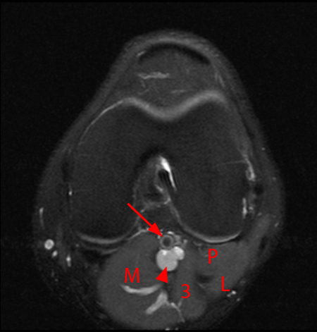
The common peroneal nerve typically courses downward within abundant fat posterior to the short head of the biceps femoris muscle and superficial to the lateral head of the gastrocnemius muscle, but. Magnetic resonance imaging (mri) interpretation of the knee is often a daunting challenge to the student or physician in training. In conclusion, we describe the normal mri anatomy of the distal biceps femoris and the relationship of this muscle with the common peroneal nerve. Cross sectional anatomy of the knee based on mri : It is the largest synovial joint in the body and allows flexion and extension of the leg as well as some rotation in the flexed position. Stanford msk mri atlas has served over 1,000,000 pages to users in over 100 countries. Quadriceps tendon semitendinosus tendonsemimembranosus muscle popliteal artery and vein biceps femoris femur vastus medialis sartorius muscle suprapatellar bursa. The smaller bone that runs alongside the tibia (fibula) and the kneecap (patella) are the other bones that make the knee joint. Medical images from an mri allow medical professionals to distinguish body tissues, including the meniscus (shock absorbers in the knee), cartilage, tendons, and ligaments. T2w axial fat sat 1. Plantaris can have variable size, but in most cases is difficult to demonstrate on routine mri studies. It is considered a vestigial muscle, and can be used as a tendon graft in reconstructive orthopedic surgery. They are attached to the femur (thighbone), tibia (shinbone), and fibula (calf bone) by fibrous tissues called ligaments.
Anterior and posterior cruciate ligaments. Both the pronounced accuracy of the mri and the high prevalence of knee disorders, makes the knee mri the most frequently ordered imaging procedure of the musculoskeletal system. This mri hip joint axial cross sectional anatomy tool is absolutely free to use. These muscles work in groups to flex extend and stabilize the knee joint. Anatomy arthrogram anatomy basic shoulder mri.
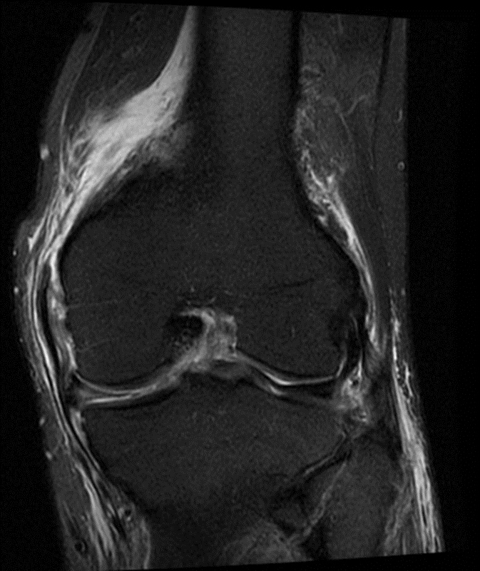
How To Read The Normal Knee Mri Kenhub from thumbor.kenhub.com Articular surface of patella and femur, condyle, epicondyle and muscles (popliteus, sartorius, gastrocnemius, semimembranous with tendos.) the images obtained were exported to jpeg from dicom data stored on the pacs (picture archiving and communicating system). Atlas of knee mri anatomy. The knee joint is a complex structure that involves bones, tendons, ligaments, muscles, and other structures for normal function. The knee joint is a synovial joint which connects the femur thigh bone the longest bone in the body to the tibia shin bone. Anatomy of the knee bones around the knee. When there is damage to one of the structures that surround the knee joint, this can lead to discomfort and disability. The knee joins the thigh bone (femur) to the shin bone (tibia). Song, uc san francisco msiv gillian lieberman md.
Anatomical structures of the lower limb (hip, thigh, knee, leg, ankle and foot) and specific regions (compartment of the lower.
The common peroneal nerve typically courses downward within abundant fat posterior to the short head of the biceps femoris muscle and superficial to the lateral head of the gastrocnemius muscle, but. Superiorly, it extends to the level of the crossing of the biceps femoris tendon, and remains superficial to fcl in this location.10 Mri knee anatomy knee sagittal anatomy free cross sectional anatomy mri knee mri diagnostic imaging : Muscle anatomy coloring sheets 12 photos of the muscle anatomy coloring sheets free. The muscles of the knee include the quadriceps, hamstrings, and the muscles of the calf. This article is based on a presentation given by david rubin and adapted for the radiology assistant by robin smithuis. Related posts of knee muscle anatomy mri muscle anatomy coloring sheets. Prescribe sagittal plane off axial images with line parallel to bony glenoid. Coronal anatomy of the knee. Atlas of knee mri anatomy. Articular surface of patella and femur, condyle, epicondyle and muscles (popliteus, sartorius, gastrocnemius, semimembranous with tendos.) find this pin and more on anatomyby radiologist.ayman almatboly. Mri wrist anatomy scroll using the mouse wheel or the arrows. When a muscle has different orientations of the tendons it means that there are different patterns of edema possible depending on the tendon injured.
Plantaris acts weakly to plantar flex the foot and flex the knee. In this presentation mri anatomy biceps femoris muscle. The knee joint is a modified hinge joint between the femur, tibia, and patella. The smaller bone that runs alongside the tibia (fibula) and the kneecap (patella) are the other bones that make the knee joint. This article is based on a presentation given by david rubin and adapted for the radiology assistant by robin smithuis.
Medial Supporting Structures Of The Knee With Emphasis On The Medial Collateral Ligament Radsource from radsource.us Assoc prof craig hacking and dr shu su et al. Injuries such as anterior cruciate ligament, meniscus and rotator cuff tears are all easily diagnosed when there is a firm understanding and knowledge of human anatomy. These motions of the knee allow the body to perform such important movements as walking, running, kicking, and jumping. Richolt j.a., jakab m., kikinis r. Anatomy basic knee mri checklist. The knee joins the thigh bone (femur) to the shin bone (tibia). Extending along the anterior surface of the thigh are the four. Anatomical structures of the lower limb (hip, thigh, knee, leg, ankle and foot) and specific regions (compartment of the lower.
It is the largest synovial joint in the body and allows flexion and extension of the leg as well as some rotation in the flexed position.
This mri knee sagittal cross sectional anatomy tool is absolutely free to use. Plantaris can have variable size, but in most cases is difficult to demonstrate on routine mri studies. Articular surface of patella and femur, condyle, epicondyle and muscles (popliteus, sartorius, gastrocnemius, semimembranous with tendos.) find this pin and more on anatomyby radiologist.ayman almatboly. Mri knee anatomy knee sagittal anatomy free cross sectional anatomy mri knee mri diagnostic imaging : Thigh muscles also protect neurovascular structures as they go through the proximal hip joint to the knee and lower leg (3). It is the largest synovial joint in the body and allows flexion and extension of the leg as well as some rotation in the flexed position. Both the pronounced accuracy of the mri and the high prevalence of knee disorders, makes the knee mri the most frequently ordered imaging procedure of the musculoskeletal system. The muscles of the knee include the quadriceps, hamstrings, and the muscles of the calf. The knee joins the thigh bone (femur) to the shin bone (tibia). The muscles of the knee include the quadriceps, hamstrings, and the muscles of the calf. Radiology imaging medical imaging subscapularis muscle shoulder anatomy bicep tendonitis mri brain shoulder rehab rotator cuff tear anatomy this. They are attached to the femur (thighbone), tibia (shinbone), and fibula (calf bone) by fibrous tissues called ligaments. In conclusion, we describe the normal mri anatomy of the distal biceps femoris and the relationship of this muscle with the common peroneal nerve.


0 Komentar